|
|
Drop Machine
prints on mycelium, 2026
Project credits:
Jara Saluena Martin - biologist
Lars Dittrich - designer
Jean Cosyn - architect
Stan Vlaminck - technician
Special thanks to Iwein Baeyens and Frank Theys
Production: VUB
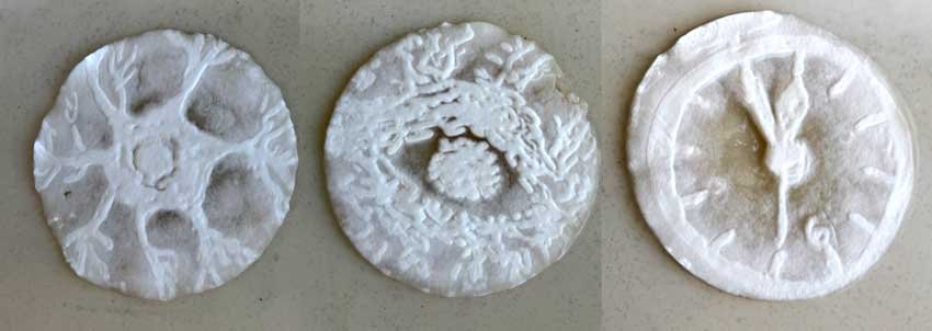
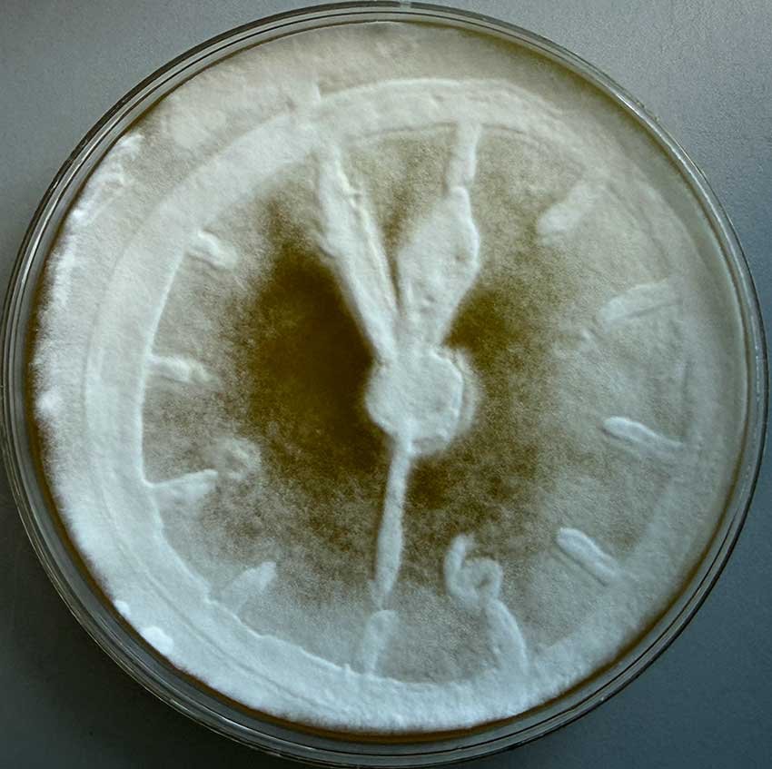
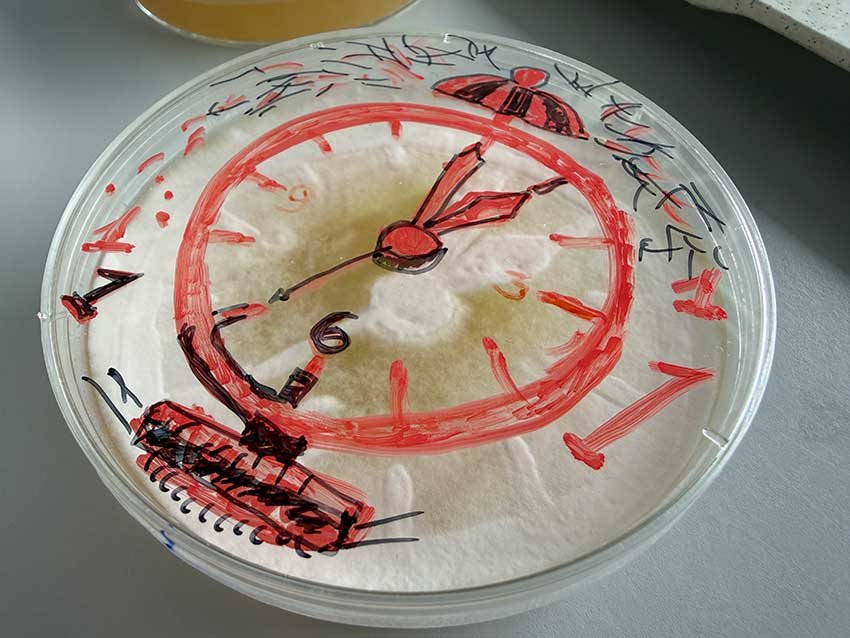
Statement:
What if a record of human thought - a photograph, a symbol, a drawn mark - could be introduced into a living organism? Not preserved within it, but offered to it?
This project begins with a photomask: a defined, legible record of a memory, idea, or representation. The mask may take the form of a photograph, a pictogram, or a drawn image. Through a photogram-like process, UV light passes through this mask and reaches a surface of growing mycelium. The light carries the image from one material condition into another.
The mycelium does not receive the image passively. It is a living, networked organism in continuous development - growing, branching, thickening, and responding to its environment. When the imprinted form meets this living surface, the result is not reproduction but negotiation: between the fixed intention of the mark and the organism’s own biological logic. This negotiation is the conceptual core of the work.
Traditional image-making tends to fix a moment. Photography has always promised permanence - light touches a surface and a trace is locked in place. Here, the receiving surface refuses that contract. The imprinted image is not stored; it enters a process. Over time, the original form may shift, fragment, blur, or be absorbed entirely into the mycelium’s expanding structure.
There is a further dimension the work cannot avoid. The organism will eventually be terminated - dried, halted, fixed - and it is in this final state that the piece enters the world as an object. Presented like a drawing or a photograph, mounted and shown, the dried mycelium carries within its structure whatever remains of the original mark: a trace, a residue, something between legible image and biological record. The artwork is finished only when the living process is stopped.
This ending is not incidental. It is part of the work’s meaning. The piece moves through a full arc - growth, negotiation, disso-lution, arrest - and what is displayed is not the process itself but its aftermath. The viewer encounters a once-living surface, now stilled, holding the faint evidence of an encounter between human intention and biological response.
This offers a different model of memory than photography usually proposes. Human memory is rarely a perfect archive. It reconstructs, fades, fragments, and resurfaces unexpectedly. The mycelium’s living response - its willingness to absorb and alter what it receives - offers a material analogy for that condition. And the dried object that results shares something with a photograph or a drawing: it is a record, but of a process rather than a moment, and the form it preserves was never entirely under the artist’s control.
Imprinting Memory does not claim that mycelium thinks, or that it stores experience in any biological sense. It uses the organism’s growth - and its arrest - as a medium through which to ask: what happens to meaning when the surface carrying it was once alive?
History:
The project “Imprinting Memory: Mycelium, Light, and the Living Image” began in early 2026, when Franck Theys invit-ed me to participate in “Eco Stories,” an exhibition exploring the relationship between art and science. The exhibition was planned in collaboration with VUB and was to be presented at the university’s PILAR gallery. For me, this invitation offered an opportunity to continue an inquiry that connects several of my previous works: how can we make visible what usually lies beyond our perception?
In my artistic practice, I often explore processes and forms of life that go unnoticed in everyday life. They may be too small to see with the naked eye, or change so gradually that their movement is difficult to perceive. A microscope, a sensor, or a data transmission system allows me to approach this world and find an artistic form through which to express it.
One of my previous projects was based on microscopic observations of living organisms and their surroundings. The result-ing images became part of an immersive projection that viewers could enter. Through this change in scale, minute move-ments and structures filled the surrounding space and became visible to us.
In a subsequent project, I worked with the fermentation of SCOBY—a symbiotic culture of bacteria and yeast used to make kombucha. I built a model of a glucose molecule using spheres with embedded LEDs. The model was connected to a sensor immersed in a container of sweetened tea and a kombucha culture.Data recording changes in the liquid during fermenta-tion were fed into a control system, altering the light patterns within the model. In this way, the process unfolding within the culture found a visible expression.
After receiving the invitation to participate in “Eco Stories,” I asked to visit the university’s Department of Microbiology (MICR). I wanted to learn about its ongoing research and explore whether the growth of biological material could itself participate in creating an image.
During my visit, I was drawn to Jara Saluena Martin’s research into the effects of different colours of light on mycelial growth. Looking at the samples, I noticed differences in their density: the mycelium appeared thicker in areas receiving more light, while it became thinner towards the edges, where there was little or almost no light. This observation gave me the idea of using the distribution of light to create a pattern within the growing material.
I proposed creating a kind of photogram on mycelium using a photomask. Light would pass through the mask at varying intensities, and the mycelium would gradually form an image as it responded to the illumination. I imagined the result as a subtle relief: differences in the thickness and density of the mycelium would remain visible after drying. In such a work, the image develops over time, and its final appearance depends on the growth of a living organism.
Jara Saluena Martin and her colleague Lars Dittrich agreed to try this method, marking the beginning of our collaboration. We first conducted trials in small Petri dishes, 9 centimetres in diameter, to test how well the photomask worked. The results were very encouraging: differences in mycelial growth made the intended image visible. Together with Lars Dittrich and VUB technicians, we then began constructing a larger box, measuring 50 × 30 centimetres, for growing the mycelium.
By the opening of the exhibition, I hope we will have between 15 and 20 small samples and three to four larger ones. To-gether, they will show both the outcome of the experiment and the variety of images that emerge through the interaction of light and growing mycelium.
I am particularly drawn to the possibility of preserving a trace of light’s influence within the material. I connect this to the way what we see leaves an imprint on our memory and continues to exist in our imagination. Here, that trace gradually takes physical form, becoming an image that holds the history of its own emergence within its surface.